HOME
ABOUT US
REFERENCES
REPRESENTATIONS
CONTACT US
TÜRKÇE
Engineering and Technical Education
PHYSICS
ELECTRONICS
COMMUNICATIONS
ELECTRICITY
ENERGY
MECHATRONICS, AUTOMATION & COMPUMECHATRONICS
MECHANICS
FLUID MECHANICS
THERMODYNAMICS & THERMOTECHNICS
PROCESS CONTROL
CHEMICAL ENGINEERING
FOOD & WATER TECHNOLOGIES
ENVIRONMENT
BIOMEDICAL ENGINEERING
Physics, Chemical, Biology Education
OPTIKASCIENCE
DATA HARVEST
ERLER-ZIMMER
Maths Education
ERLER-ZIMMER
Anatomical Models / Skeleton Models / Skulls
1. OPTIKASCIENCE
1.1. Section 01 - Kits
1.1.1. Physics Laboratory Sets
1.1.2. Basic Kits
1.1.3. Advanced Kits
1.2. Section 02 - Physics
1.2.1. Statics of solids
1.2.2. Dynamics
1.2.3. Translational motion
1.2.4. Rotational motion
1.2.5. Oscillatory motion
1.2.6. Inertia- Collisions - Two-dimension motion
1.2.7. Liquids
1.2.8. Gases and vacuum
1.2.9. Wave's propagation
1.2.10. Sound Waves
1.2.11. Molecular aspect of matter
1.2.12. Temperature and Heat
1.2.13. Geometrical Optics
1.2.14. Wave Optics
1.2.15. Optical Benches
1.2.16. Electrostatics
1.2.17. Electrical conduction
1.2.18. Magnetism and electromagnetism
1.2.19. Atomic Physics
1.3. Section 03 - Technique and Energy
1.3.1. Energy conversions
1.3.2. Renewable energies
1.4. Section 04 - Microscopy
1.4.1. On-field microscopy kits
1.4.2. Biological microscopes
1.4.3. Stereomicroscopes
1.4.4. Multimedia system
1.4.5. Microscopy accessories
1.4.6. Optical magnifiers
1.4.7. Prepared slides for microscopy
1.5. Section 05 - Biology
1.5.1. Botany
1.5.2. Zoology
1.5.3. Experiments on human beings
1.5.4. Human anatomy and DNA models
1.6. Section 06 - Ecology
1.6.1. Kit for environmental analysis
1.6.2. Items for sample's collection
1.6.3. Stations for the detection of air pollution
1.6.4. Digital instruments
1.7. Section 07 - Meterology
1.7.1. Instruments and weather stations
1.8. Section 08 - Astronomy and Earth Science
1.8.1. Rocks, fossils and minerals
1.8.2. Geological models
1.8.3. The Earth and the solar system
1.9. Section 09 – Chemistry
1.9.1. Periodic table of elements
1.9.2. Molecular models and atomic models
1.9.3. PH-meters
1.9.4. Refractometry
1.9.5. Polarimetry
1.9.6. Spectroscopy
1.9.7. Electrology
1.10. Section 10 – On-Line Science
1.10.1. Interfaces
1.10.2. MBL Sensors
1.10.3. USB Sensor
1.11. Section 11 – Drawing and Mathematics
1.11.1. Drawing
1.11.2. Enumeration
1.11.3. Logics
1.11.4. Fractions and percentages
1.11.5. Geometry
1.11.6. Mathematics on magnetic blackboard
1.12. Section 12 – Measurement Instruments
1.12.1. Lengths and angles
1.12.2. Volumes/Time intervals
1.12.3. Temperature
1.12.4. Density/Forces, weights and masses
1.12.5. Electrical devices
1.13. Section 13 – Lab Tools
1.13.1. Items and instruments
1.13.2. Electrical power sources
2. DATA HARVEST
2.1. Data Logging
2.1.1. Data Loggers
2.1.1.1. Data Logging Range
2.1.2. Sensors
2.1.2.1. Sensor Range
2.1.3. Apps
2.1.3.1. Cross-platform software
2.1.4. Accesories
2.1.4.1. All Accessories
2.2. Dynamics
2.2.1. Dynamics System
2.2.2. Software
2.2.2.1. Cross-platform software
2.2.3. Accessories
2.2.3.1. All Accessories
2.3. Teaching
2.3.1. Teaching Packs
2.3.2. Teaching Metarials
2.4. More Science
2.4.1. Smart Microscopes
2.4.2. Genecon Hand-Held Dynamo
2.4.3. Renewable Energy
2.5. Product Brochures
2.5.1. Primary Teaching Brochure
2.5.2. Secondary Teaching Brochure
3. ERLER-ZIMMER
3.1. Anatomical Models
3.1.1. Skeleton Models
3.1.1.1. Pelvises
3.1.1.2. Limbs
3.1.1.3. Full Body
3.1.1.4. Skulls
3.1.1.5. Spines
3.1.1.6. Bones
3.1.2. Organs & Structures
3.1.2.1. Respiratory System
3.1.2.2. Brain & Nerves
3.1.2.3. Urinary System
3.1.2.4. Skin-Hair-Nail
3.1.2.5. Head Models
3.1.2.6. Circulatory System - Blood Vessels
3.1.2.7. Circulatory System - Hearts
3.1.2.8. Circulatory System - Circulation
3.1.2.9. Liver
3.1.2.10. Muscular Models
3.1.2.11. Kidneys
3.1.2.12. Ears
3.1.2.13. Torsos
3.1.2.14. Digestive System
3.1.2.15. Teeth
3.2. 3D Anatomy Series
3.3. Medical Simulators
3.3.1. Auscultation
3.3.2. Endoscopy
3.3.3. Gynaecology
3.3.4. Skin Suture
3.3.5. Injection
3.3.6. Nursing
3.3.6.1. Chronic Wounds
3.3.6.2. Diabetes
3.3.6.3. Catheterisation
3.3.6.4. Nursing Dolls - Baby & Child Dolls
3.3.6.5. Nursing Dolls - Adult Dolls
3.3.6.6. Stoma Care
3.3.7. Cancer Prevention
3.3.8. Emergency
3.3.8.1. Airway
3.3.8.2. CPR - Baby & Child Dolls
3.3.8.3. CPR - Adult Dolls
3.3.8.4. All Dolls - Baby & Child Dolls
3.3.8.5. All Dolls - Adult Dolls
3.3.8.6. Rescue
3.3.8.7. Thorax
3.3.8.8. Trauma Simulation
3.3.9. X-RAY / CT
3.3.10. Pregnancy & Birth
3.3.11. Ultrasound
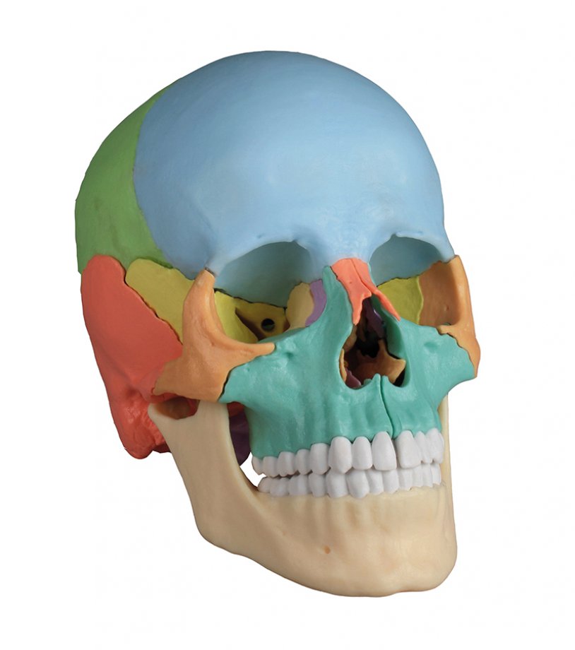
Osteopathic Skull Model, 22 part, didactical version
This fascinating model of an average European adult skull can be disassembled into 22 single bones. During development of this model one of the main targets was to make the model easy to assemble and dismantle. Stable parts with convenient magnet connections make handling of the product a child‘s play. The detailed bones do not need any complicated pins to be stuck into holes, they almost slide into position, guided by realistic bone sutures and held by strong magnets. Due to the very good anatomy and the easy handling this skull is the ideal training aid for osteopathy. This version is perfect for visual ...
Osteopathic Skull Model, 22 part, anatomical version
This fascinating model of an average European adult skull can be disassembled into 22 single bones. During development of this model one of the main targets was to make the model easy to assemble and dismantle. Stable parts with convenient magnet connections make handling of the product a child‘s play. The detailed bones do not need any complicated pins to be stuck into holes, they almost slide into position, guided by realistic bone sutures and held by strong magnets. The perfect tool for Osteopaths. The following bones are represented: - Parietal bone left and right - Occipital bone - Temporal bone le...
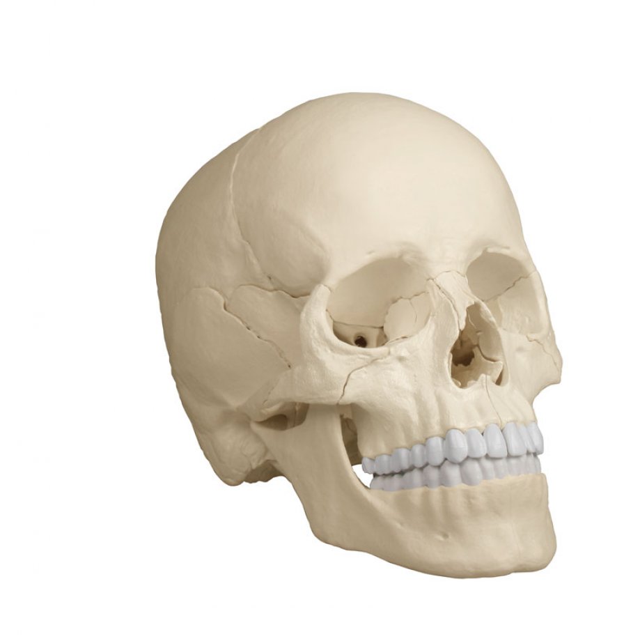
Skull model, 3 parts
Developing this lifelike reproduction of a human skull we have used the latest technology to digitalize a real human skull and idealized it under the aspects of medical education. This means the skull was adjusted to be anatomically ideal, all anatomical details and structures are present and correspond to anatomical teaching. The three part model consists of skull base, skull cap and lower jaw. The teeth correspond with real dentition concerning position and embrasure. The lower jaw is movably mounted and can be removed. The skull cap is aligned with the skull base using metal pins and held by strong magnets. Du...
Dental skull, 4-part
The teeth in the upper and lower jaw can be extracted and reinserted. The mandible is open and shows roots, spongiosa, nerve canal and an impacted wisdom tooth. The mandible is removable. Size: 22 x 13 x 17 cm, weight: 0.7 kg...
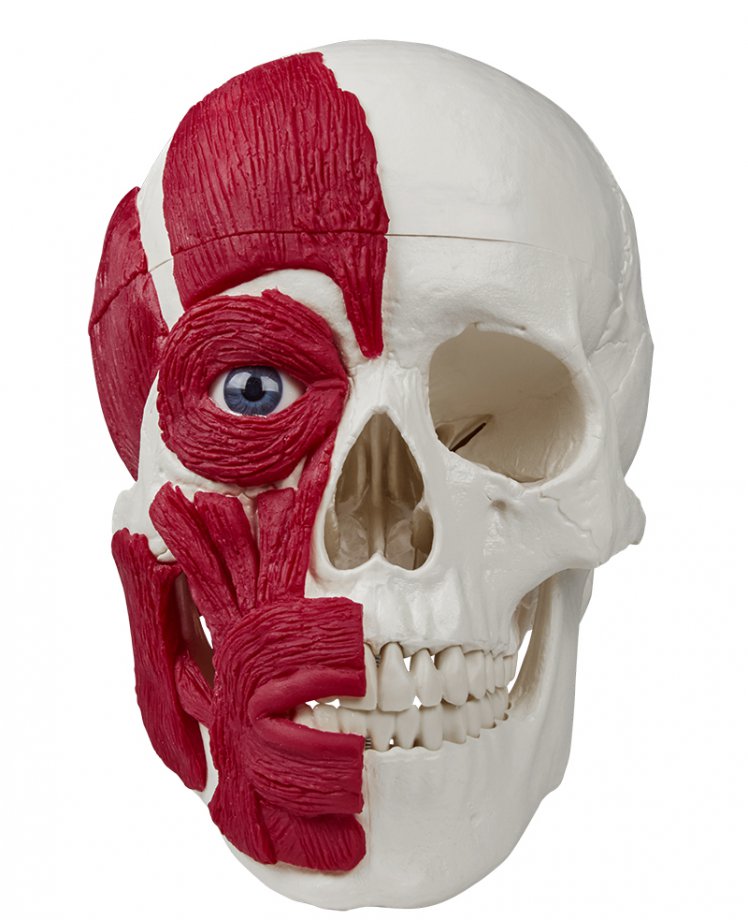
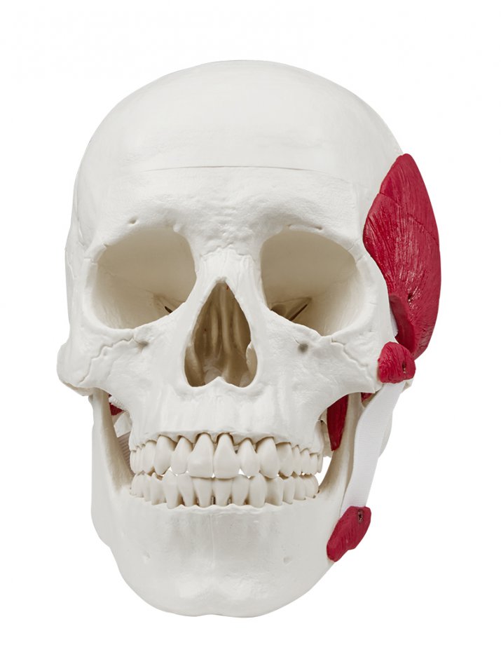
Skull with musculature
This fascinating model shows a human skull with the most important head muscles on one half. The skull is similar to the article 4500, the mandible is unmovable. The skull cap can be opened and removed. Size: 18 x 19 x 12 cm, weight: 0.8 kg...
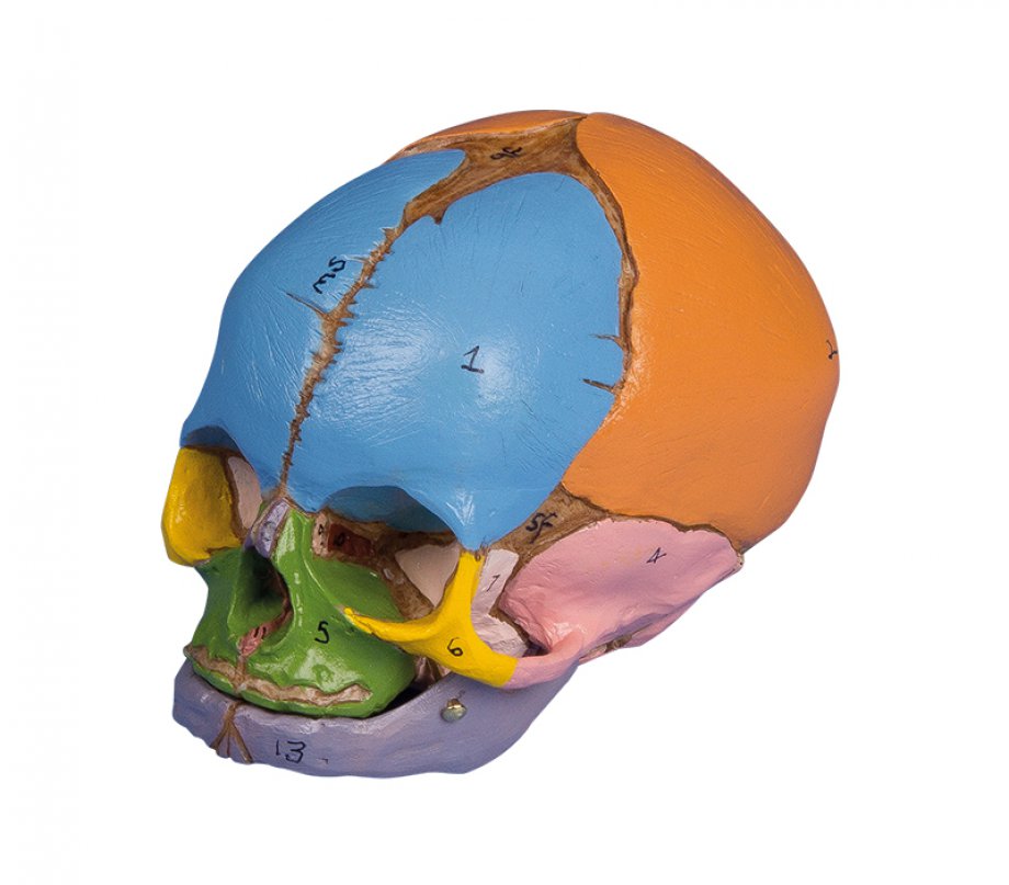
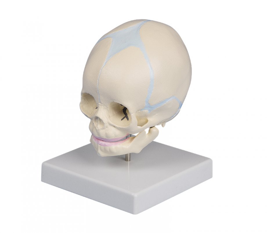
Didactic Foetal Skull, 38. week
A very realistic model of a 38-week foetal skull manufactured from our unique bone-like material. The separate skull bones are clearly shown and meticulous moulding distinguishes such detail as sutures, fontanelles and the external auditory meatus. The main bones of the skull coloured for easy identification. Supplied with key card....
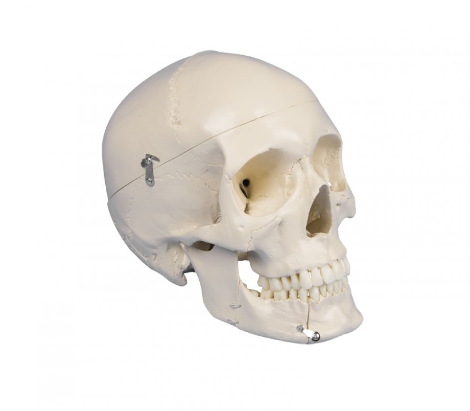
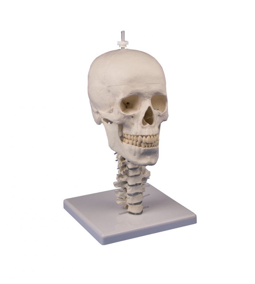
Skull model, 3 parts with skull stand cervical spine
Developing this lifelike reproduction of a human skull we have used the latest technology to digitalize a real human skull and idealized it under the aspects of medical education. This means the skull was adjusted to be anatomically ideal, all anatomical details and structures are present and correspond to anatomical teaching. The skull consists of skull base, skull cap and lower jaw. The teeth correspond with real dentition concerning position and embrasure. The lower jaw is movably mounted and can be removed. The skull cap is aligned with the skull base using metal pins and held by strong magnets. Due to this t...
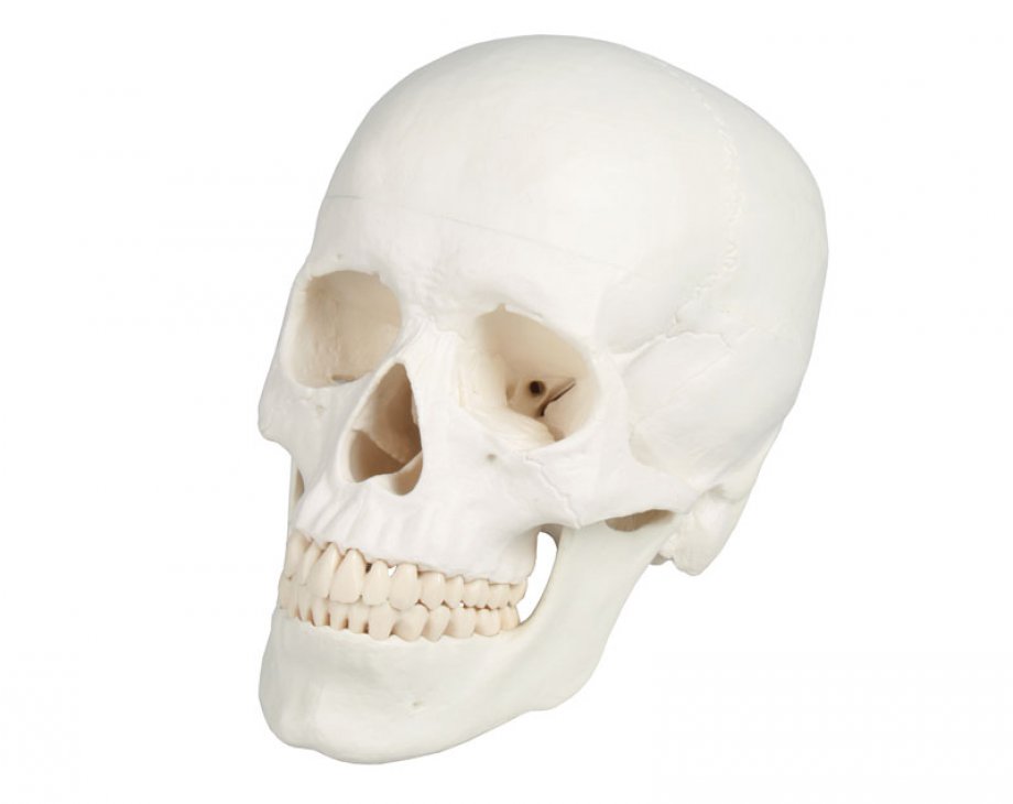
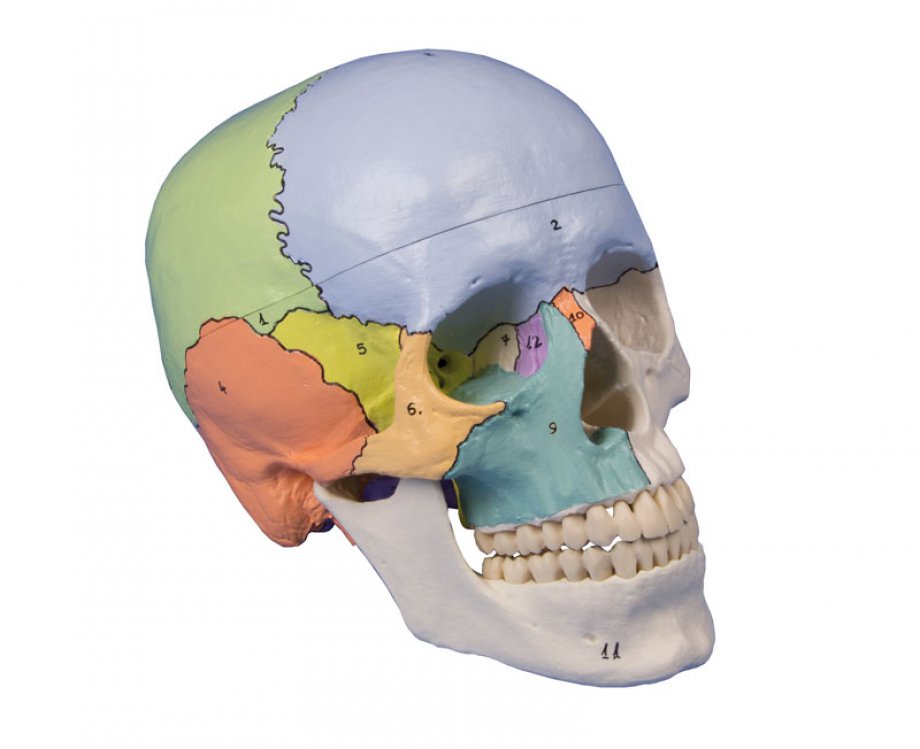
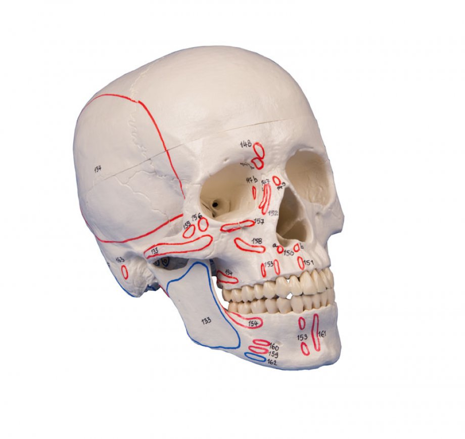
Skull model, 3-part, didactical painted
Skull model like ref.no. 4500, additionally with didactical painting of individual bones on one side of the skull. The bones are numbered referring to the included nomenclature. Size: 18 x 19 x 12 cm, weight: 0.7 kg...
Skull model, 3-part, with muscle marking
Skull model like ref.no. 4500, but additionally with marking of muscle insertions and origins. With nomenclature. Size: 18 x 19 x 12 cm, weight: 0.7 kg...
Skull with masticatory muscles, 3-part
The masticatory muscles (masseter muscle, temporal muscle, pterygoideus medialis and lateralis muscles) are represented in the form of elastic bands. Using this model, it is possible to demonstrate the function of the masticatory muscles with closure of the mandible, induction of opening of the mandible and lateral and forward displacement of the mandible. The the skull cap is removable, the mandible is moveable. Size: 23 x 16.5 x 17 cm, Weight: 0.8 kg...
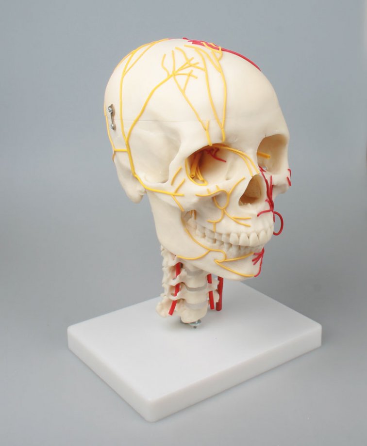
Neurovascular Skull
This model shows a life size skull with seven cervical vertebrae. The arteries are shown on one side and nerves on the other. Removing the vault exposes the main nerves and arteries inside the skull. The 12 cranial nerves and their branches are also shown. Life size....
Fetus skull, 30. week
This model represents a human foetal skull in the 30. week of pregnancy. Size: 12 x 12 x 27 cm, weight: 0.4 kg...
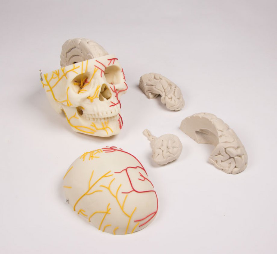
Neurovascular skull with brain
The three part skull shows the main skull nerves and arteries. The brain has 8 parts and is made of soft, tissue like material. Size: 17.5 x 16.5 x 22 cm...
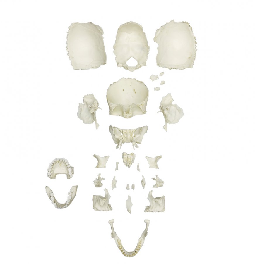
Disarticulated Human Medical Study Skull
In order to develop this skull properly, the advice of educators, anatomists and surgeons was solicited.Following an evaluation from Dr. Noel Boaz, Professor of Anatomy: „This skull is ideal for osteological teaching. It allows the student to appreciate the individual bones, the sutures between the bones, and the foramina of the skull. All foramina are bristle-patent, making student exploration of perforating cranial nerve branches and blood vessels possible.For the medical or dental student this skull is an excellent companion to head-and-neck dissection. For the physical anthropology student this skull i...
Disarticulated Human Fetal Skull, Full Term
This disarticulated skull is from a full term (10 lunar months) fetus. It was part of a medical examiner‘s comparative pathology collection before going to the Maxwell Museum and is remarkable in its completeness. It was possible to reproduce every bone, small and large, in precise detail. A perfect specimen for study in forensic anthropology, physical anthropology and anatomy....